A 24-year-old female patient with an incidental finding on chest radiograph, performed in an asymptomatic patient because of a routine medical check-up. The following chest radiographs were acquired (Fig. 1).
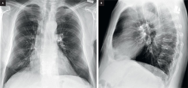
Figure 1. a, PA chest x-ray, b, lateral chest x-ray.
Part 1. Chest Radiographs
Questions for discussion
1. What is the abnormality on the chest radiographs?
2. What are the differential considerations for the findings?
Explanation
Chest radiograph shows a subtle opacity over the right hilum and paratracheal region on the frontal chest radiograph (Fig. 2).
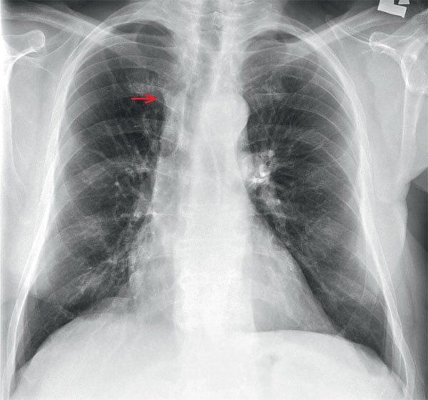
Figure 2. Chest x-ray with the right hilar opacity (arrow).
Differential consideration would include: abnormal lymph node, distended azygous vein, and a small para-tracheal lesions.
Part 2. CT scan
The patient subsequently underwent a chest CT.
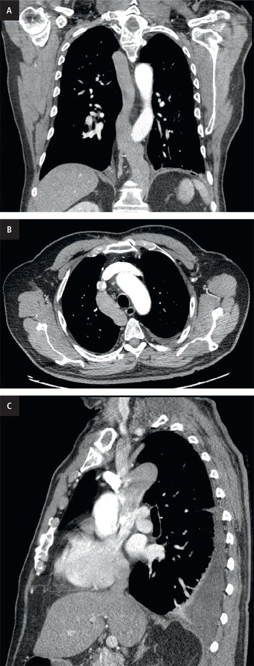
Ryc. 3. CT scan. a, coronal image, b, axial image, c, sagittal image.
Questions for discussion
1. What is the abnormality?
2. Are there any associated conditions to be aware of?
Explanation
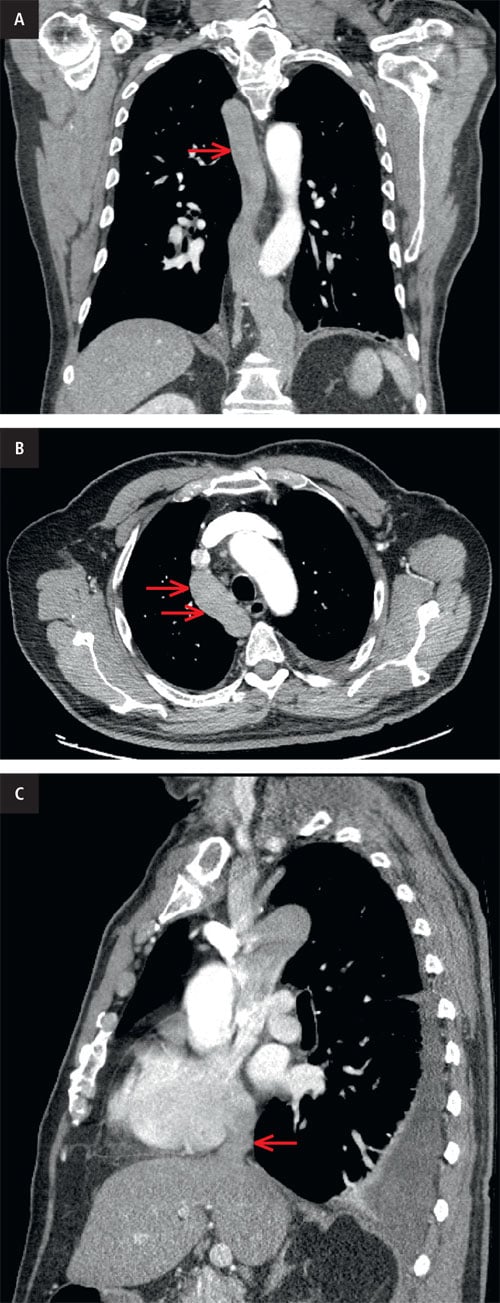
Figure 4. CT scan. The coronal image (A) clearly shows the dilated azygos vein (arrow) corresponding with the right hilar opacity originally observed on chest radiograph. The axial image (B) also clearly shows the dilated azygos vein which drains into the superior vena cava (arrows). The sagittal image (C) shows the hepatic veins draining directly into the right atrium with no inferior vena cava draining into the right atrium (arrow).
CT confirms the presence of an azygos continuation of the IVC.
There is more clear delineation of the variant anatomy with interruption of the infrahepatic suprarenal IVC due to an absent fusion of the infrahepatic and suprarenal IVC during embryogensis. Blood instead drains through a dilated azygos vein. Azygous continuation of the IVC is an uncommon vascular anomaly representing an anatomical variant. It is important to recognize as the findings can be mistaken for a right paratracheal mass or adenopathy. On its own, azygous continuation of the IVC is most often an incidental finding with little clinical consequence. On chest radiographs, it could present as an apparent abnormality in the right para-tracheal region.
Diagnosis
Azygos continuation of the inferior vena cava
Azygous continuation of the IVC most often occurs in isolation and patients are asymptomatic. It is important to recognize as it can have implications for patients undergoing endovascular or cardiopulmonary bypass procedures.
You can find more interesting cases in the app "Discover Radiology: Chest x-ray interpretation". Only $6.99!
See more than meets the eye!
References
1. Bass J.E., Redwine M.D., Kramer L.A., Huynh P.T., Harris J.H. Jr.: Spectrum of congenital anomalies of the inferior vena cava: cross-sectional imaging findings. Radiographics. 2000; 20: 639–52.2. Ghandour A., Partovi S., Karuppasamy K., Rajiah P.: Congenital anomalies of the IVC-embryological perspective and clinical relevance. Cardiovasc Diagn Ther. 2016; 6: 482–492.