Definition, Etiology, Clinical FeaturesTop
Subcutaneous emphysema refers to the presence of air in the subcutaneous tissue. Most frequently it develops as a result of air leakage from a pneumothorax or pneumomediastinum into the subcutaneous tissue of the neck (less commonly the chest, head, or abdomen). On rare occasions it is caused by gastrointestinal (GI) perforation below the navel.
Signs and symptoms: Discomfort in the neck and chest, crackles on compression of the neck and supraclavicular region, features of pneumothorax or pneumomediastinum.
DiagnosisTop
Chest radiographs reveal air in the subcutaneous tissue of the neck and chest (Figure 1). This is accompanied by radiologic features of pneumothorax, pneumomediastinum, or perforation of the GI tract (air under a hemidiaphragm seen on plain abdominal radiographs). Differential diagnosis includes CO2 insufflation for laparoscopic surgery.
TreatmentTop
In patients with subcutaneous emphysema associated with pneumomediastinum and not caused by esophageal, tracheal, or bronchial perforation, medical management (monitoring) is sufficient. Patients should be closely observed because the progression of subcutaneous emphysema towards the neck tissue can lead to upper airway obstruction. Subcutaneous emphysema coexisting with a pneumothorax requires the use of chest drainage to release the pneumothorax. Subcutaneous emphysema associated with the GI tract or airway perforation is an indication for urgent surgical intervention.
FiguresTop
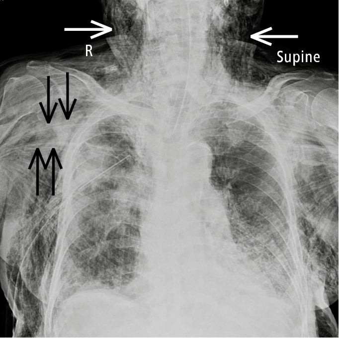
Figure 1.37-1. Anteroposterior (AP) chest radiography of a patient with a right-sided pneumothorax (with chest drain insertion) complicated by extensive subcutaneous emphysema. A large amount of air spreads into the subcutaneous tissue around the neck (white arrows) and pectoralis major muscles (black arrows).
 English
 Español
 українська