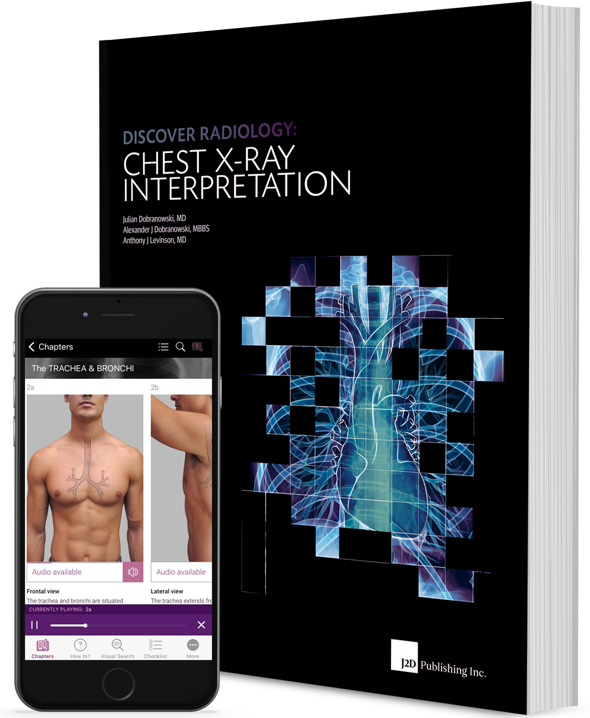
Now available in the form of a well-designed mobile application that facilitates learning and revision through features such as easy navigation, notes, quick search, and the author’s audio commentary to the How to guides.
Price – only $6.99
Structure
Section I
provides an overview of how x-rays are used to produce an image of the chest. The section is divided into 5 chapters that examine the building blocks necessary for chest x-ray interpretation. The chapters review x-ray image production and the general concepts related to image appearance. With this background information you will then be introduced to the key components of the interpretation process.
Section II
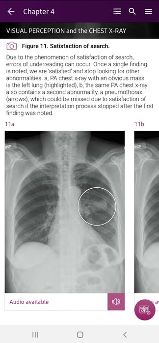
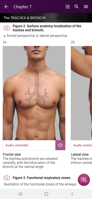
starts with a description of the radiological zones. The concept of radiological zones is introduced to give you a starting point in the understanding of the radiological anatomy of the chest. The chapters review in detail the radiological anatomy of specific anatomical structures, also provide examples of how the x-ray image can change due to pathology. The final chapter explains how the individual structures come together to form the radiological image. The focus of this section is on the anatomy of the normal PA and lateral chest x-ray - the radiological anatomy - and the basics of how pathology can alter this normal radiological appearance.
Section III
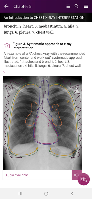
starts with a chapter on how to interpret the chest x-ray; this is where you begin to put your new knowledge into practice. Now that you know what to look for, we’ll help you do so in a systematic way to ensure the accurate identification of pathology. Subsequent chapters show you how to apply this knowledge to more complex chest x-rays. In this section you will learn further interpretive skills such as how to accurately localize pathology and how to use the digital technologies with the interpretive process. Finally, you will learn when it is appropriate to order a chest x-ray.
Special content
How To
Step by step guides, with annotated x-rays, to illustrate key skills needed to confidentially interpret chest x-ray.
Visual Search
Numbered visual guides to illustrate the sequential checks that should be performed in a visual search of given anatomical structure or radiological zone on a chest x-ray.
Radiological Checklist
Illustrated list of items that should be evaluated for a given anatomical structure or radiological zone, in the process of interpreting a chest x-ray.
Radiological Anatomy
Descriptions of various anatomical structures as they would appear on PA and lateral chest x-rays.
Case Study
Practical and Clinical Case Studies collected from all Chapters help check current skills.
Pathology
Images, divided according to specific anatomical structures and regions found within the thorax, help identify kind of pathology.
Application Features
- Instantly access Case Studies, Checklists, How to, Radiological Anatomy through the Quick List.
- Take your own notes in the app and access them whenever needed.
- Add chapters and sections to your favorites.
- Highlight most important fragments.
- Listen to the author’s commentary on How To sections.