Author Information
Julian Dobranowski, MD, FRCPC FCAR
Professor Radiology, Chair of the Department of Radiology McMaster University, Hamilton, Ontario
Alexander J Dobranowski, MBBS
Anthony J Levinson, MD, FRCPC, MSc, MA
Director, Division of e-Learning Innovation and machealth.ca John Evans Chair in Educational Research Associate Professor, Faculty of Health Sciences McMaster University, Hamilton, Ontario
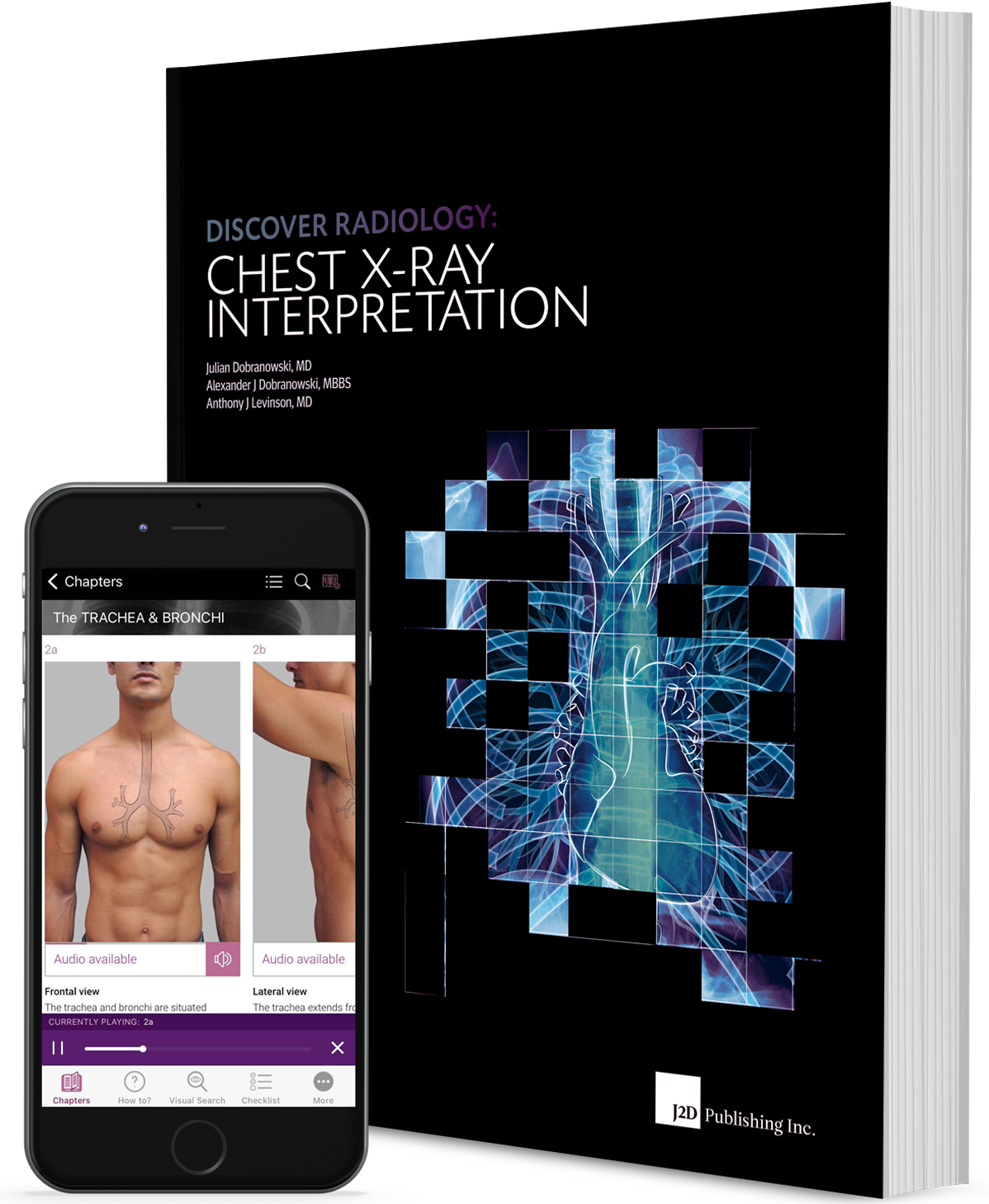
Discovery Radiology: Chest X-Ray Interpretation
Is the first in a series of radiology textbooks concentrating on the teaching of x-ray interpretation through a focus on anatomy, radiological anatomy, concepts of pathology, differential diagnosis and important concepts and competencies. Use this book to systematically develop foundational skills in chest x-ray interpretation, as well as a reference tool with job aides to continue to improve your knowledge and skills in the clinical setting. This book is your ‘one-stop-shop’ to learn, master, and achieve chest x-ray interpretative success.
Learning to interpret x-ray images is an intellectual journey. A journey to understand the truths the x-ray is trying to show us and the truths the x-ray is hiding.
During the journey through the book you will review and understand:
- the radiological anatomy of the chest
- the importance of the lateral chest x-ray
- concepts of radiological pathology
- the importance of the differential diagnosis
- appropriate ordering of radiological examinations.
How to use this book
This book is written to allow maximum flexibility during the learning experience.
It is recommended that first time around the learner experiences the book systematically from beginning to end. This is because each chapter builds on the previous and each section enhances the next.
The chapters form building blocks each playing an important role in the understanding of the interpretive process.
The breakdown of the Sections in the book is as follows:
Section I
- What is a chest x-ray?
- What is the chest x-ray image?
- What is the interpretive process?
The section is divided into 5 chapters that examine the building blocks necessary for chest x-ray interpretation. The chapters review x-ray image production and the general concepts related to image appearance. With this background information you will then be introduced to the key components of the interpretation process.
Section II
- Radiological anatomy
- How does pathology show on an x-ray?
The concept of radiological zones is introduced to give you a starting point in the understanding of the radiological anatomy of the chest. The chapters review in detail the radiological anatomy of specific anatomical structures, also provide examples of how the x-ray image can change due to pathology. The final chapter explains how the individual structures come together to form the radiological image.
The focus of this section is on the anatomy of the normal PA and lateral chest x-ray - the radiological anatomy - and the basics of how pathology can alter this normal radiological appearance.
Section III
- Applying the interpretive process
Now that you know what to look for, we’ll help you do so in a systematic way to ensure the accurate identification of pathology. Subsequent chapters show you how to apply this knowledge to more complex chest x-rays. In this section you will learn further interpretive skills such as how to accurately localize pathology and how to use the digital technologies with the interpretive process. Finally, you will learn when it is appropriate to order a chest x-ray.
How To
Step by step guides, with annotated x-rays, to illustrate key skills needed to confidently interpret chest x-rays.Visual Search
Numbered visual guides to illustrate the sequential checks that should be performed in a visual search of a given anatomical structure or radiological zone on a chest x-ray.Radiological Checklist
Illustrated lists of items that should be evaluated for a given anatomical structure or radiological zone, in the process of interpreting a chest x-ray.Additional Information
Information that the authors deem relevant for a deeper understanding of the content, but that is not critical for your understanding of chest x-ray interpretation.Frequency Of Visualization
Statistics of the frequency that important structures can be identified on x-rays.Important Point
Information that the authors deem to be extremely important to note about chest x-ray interpretation.Radiological Anatomy
Descriptions of various anatomical structures as they would appear on PA and lateral chest x-rays. This infomation is further highlighted by its presentation on black pages.What makes this book different?
- Conceptual approach to x-ray and grayscale interpretation
- Evidence-informed content and instructional design
- Mapped to specific competencies
- Performance support tools, including over 50 job aides
- Learner-centered, written with student input
Softcover
Textbook 871 pages
Trim Size 11.8110 X 9.0551 in (300 mm X 230 mm)
Publisher J2D Publishing Inc.
ISBN 978-0-9918753-0-6
Copyright 2014
Special price $89.99 (regular price 95.99) + applicable taxes and shipping