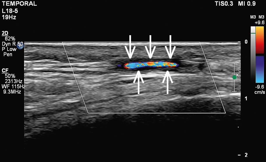
Doppler ultrasonography of the temporal artery with features of inflammation. A visible halo sign: homogenous hypoechoic wall thickening clearly separated from the arterial lumen seen on transverse and longitudinal views, typically concentric (arrows).