Figure 031_1907.

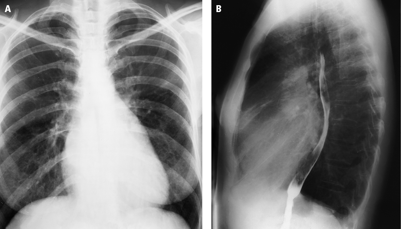
Posteroanterior (PA; A) and lateral (B) chest radiography: so-called mitral configuration of the heart. Dilatation of the main pulmonary artery, right ventricular outflow tract, and left atrium lead to straightening of the left cardiac border (mitralization) in the PA view (sometimes referred to as the mitral configuration of the heart). The lateral view shows posterior displacement of the esophagus by the left atrium. Figure courtesy of Dr Olgierd Kapuściński.

Figure 031_1907.  Posteroanterior (PA;   A  ) and lateral (  B  ) chest radiography: so-called mitral configuration of the heart. Dilatation of the main pulmonary artery, right ventricular outflow tract, and left atrium lead to straightening of the left cardiac border (mitralization) in the PA view (sometimes referred to as the mitral configuration of the heart). The lateral view shows posterior displacement of the esophagus by the left atrium.  Figure courtesy of Dr Olgierd Kapuściński.