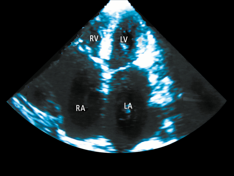
Echocardiography (apical 4-chamber view) of a patient with restrictive cardiomyopathy showing significant enlargement of both atriums (LA, left atrium; RA, right atrium). The left ventricle (LV) and right ventricle (RV) are normal sized.