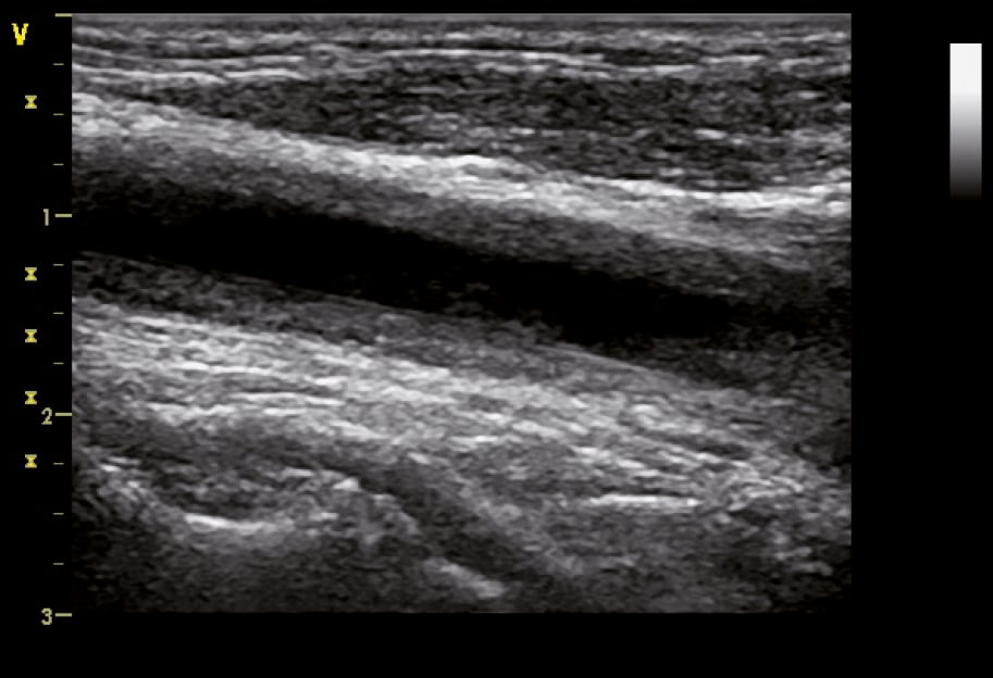
B-mode ultrasonography of the common carotid artery in a 32-year-old woman with Takayasu arteritis showing homogenous hypoechoic wall thickening (wall echogenicity increases in late disease) clearly separated from the arterial lumen, which is typical of vasculitis. Figure courtesy of Dr Leszek Masłowski.