Figure 031_6082.

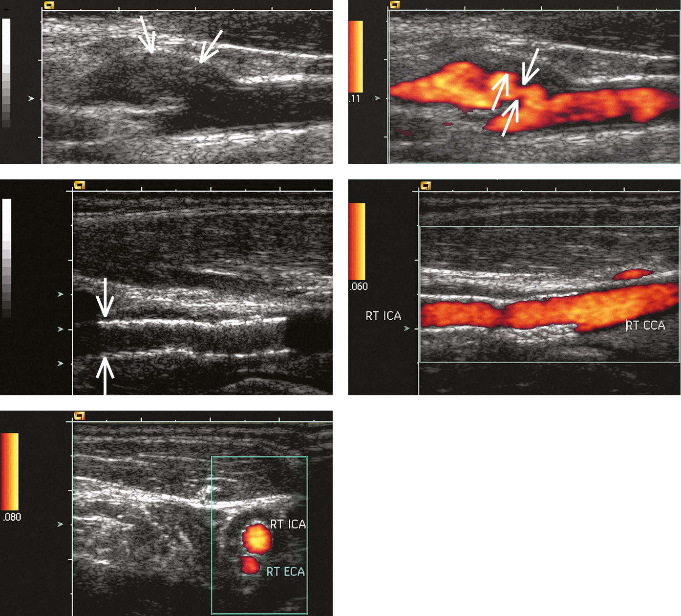
Carotid ultrasonography: A, B-mode longitudinal image of the carotid sinus and proximal segment of the right internal carotid artery (ICA). A poorly demarcated parietal lesion is seen in the proximal ICA segment, most likely atherosclerotic plaque (arrows); B, power Doppler ultrasonography shows arterial stenosis and deep ulceration (thick arrow) of the stenotic lesion (thin arrows; arterial lumen marked with color); C, D, a stent graft visible at the bifurcation of the common carotid artery, wide arterial lumen; E, stent grafts seen on the transverse image right above the common carotid artery bifurcation into the right ICA and right external carotid artery (ECA), normal arterial lumen. Figure courtesy of Dr Leszek Masłowski.

Figure 031_6082.  Carotid ultrasonography:  A , B-mode longitudinal image of the carotid sinus and proximal segment of the right internal carotid artery (ICA). A poorly demarcated parietal lesion is seen in the proximal ICA segment, most likely atherosclerotic plaque (arrows);  B , power Doppler ultrasonography shows arterial stenosis and deep ulceration (thick arrow) of the stenotic lesion (thin arrows; arterial lumen marked with color);  C ,  D , a stent graft visible at the bifurcation of the common carotid artery, wide arterial lumen;  E , stent grafts seen on the transverse image right above the common carotid artery bifurcation into the right ICA and right external carotid artery (ECA), normal arterial lumen.  Figure courtesy of Dr Leszek Masłowski.