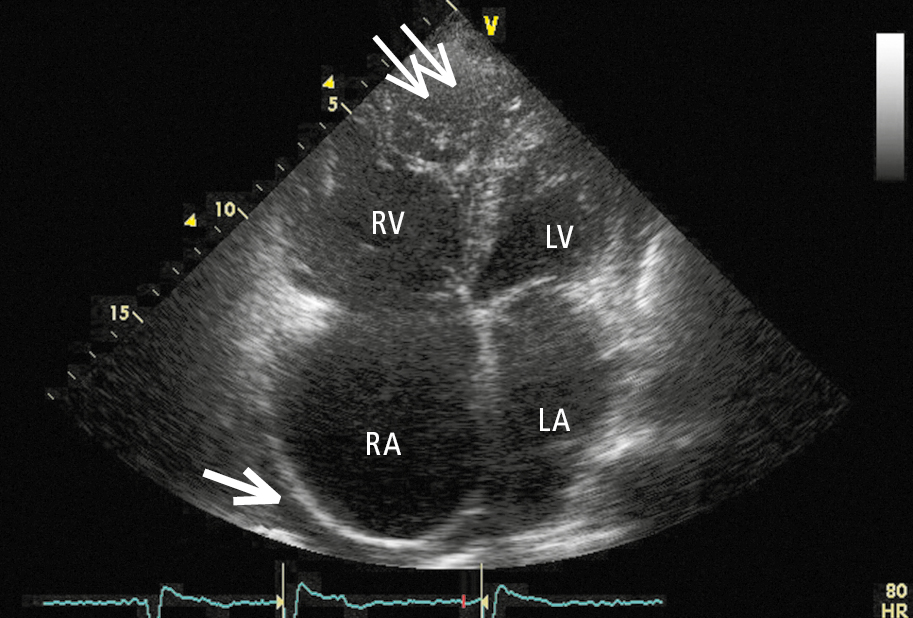
Transthoracic echocardiography (TTE) of a patient with arrhythmogenic right ventricular cardiomyopathy (ARVC). A modified apical 4-chamber view showing diffuse right ventricular injury, ample trabeculation at the apex (thin arrows), significant right atrial dilatation, and separation of pericardial layers due to pericardial fluid accumulating behind the right atrium (thick arrow). LA, left atrium; LV, left ventricle; RA, right atrium; RV, right ventricle. Figure courtesy of Dr Marek Konka.