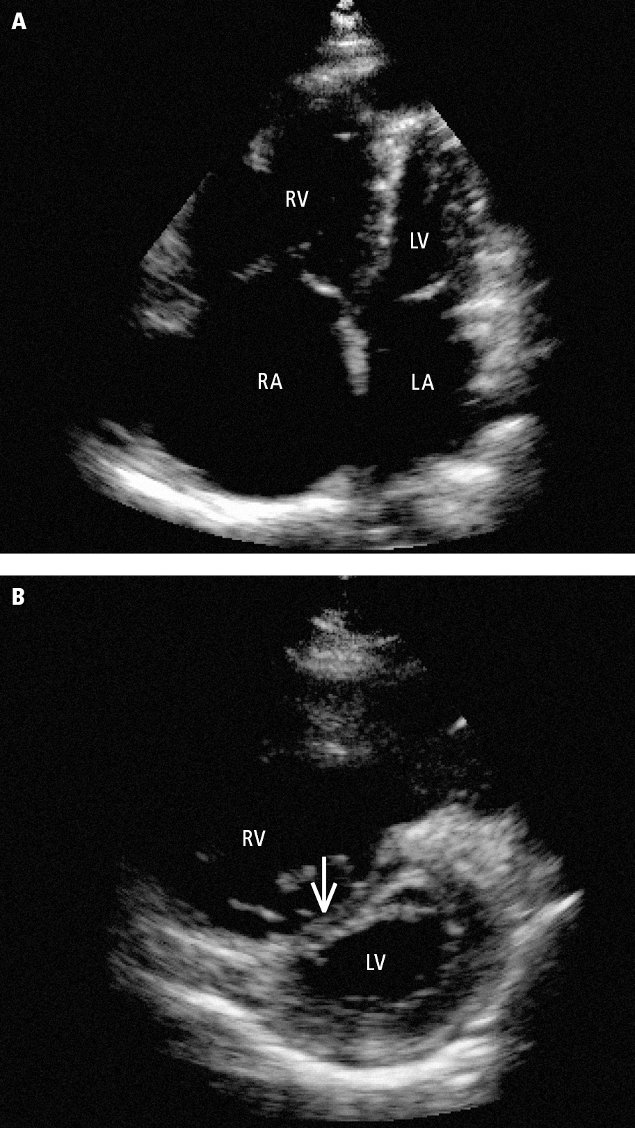
Transthoracic echocardiography (TTE) of right ventricular overload in a patient with pulmonary embolism: A, an enlarged right ventricle dominating the left ventricle seen in the apical 4-chamber view; B, flattened interventricular septum (arrow) seen in the parasternal short-axis view. LA, left atrium; LV, left ventricle; RA, right atrium RV; right ventricle.