Figure 031_7563.

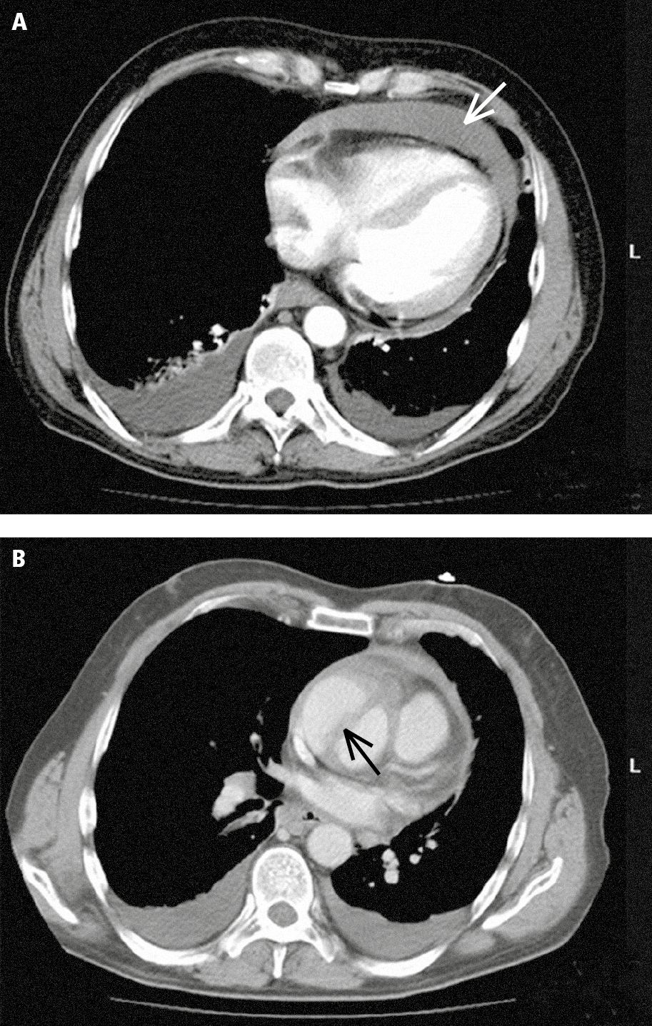
Multislice computed tomography (CT) in a patient with acute ascending aortic dissection: A, pericardial effusion (white arrow) and pleural effusion (red arrows); B, dissected ascending aorta (the arrow marks the dissection flap). Figure courtesy of Dr Jerzy Walecki.

Figure 031_7563.  Multislice computed tomography (CT) in a patient with acute ascending aortic dissection:  A , pericardial effusion (white arrow) and pleural effusion (red arrows);  B , dissected ascending aorta (the arrow marks the dissection flap).  Figure courtesy of Dr   Jerzy Walecki .