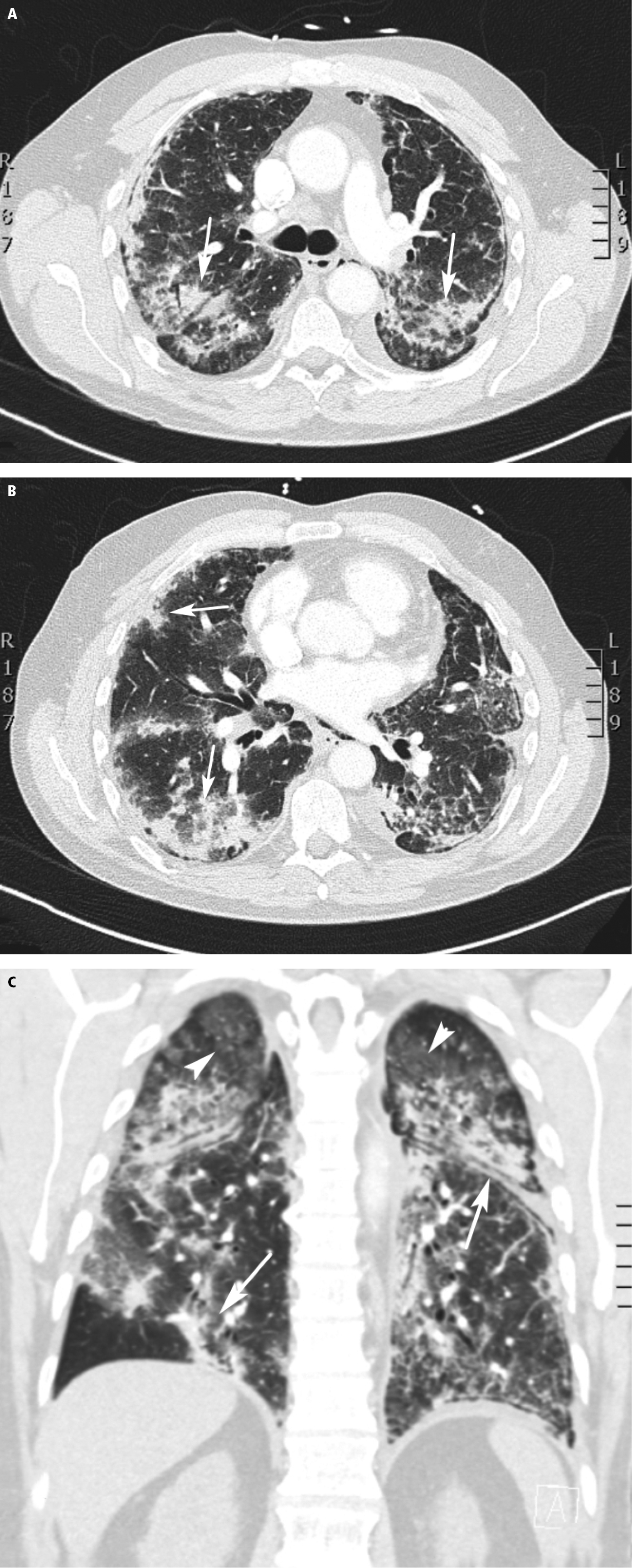
Computed tomography (CT) of a 65-year-old male patient (case #2) shows areas of consolidation (arrows) in axial (A, B) and coronal (C) planes. Rounded ground-glass opacities (arrowhead) and consolidation (arrows) are visible.