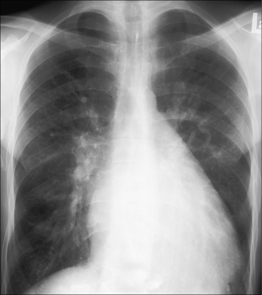
Chest radiography of a patient with ventricular septal defect (VSD): dilatation of the left ventricle, main pulmonary artery, and pulmonary arteries; features of increased pulmonary blood flow; narrow ascending aorta. Figure courtesy of Dr Piotr Hoffman.