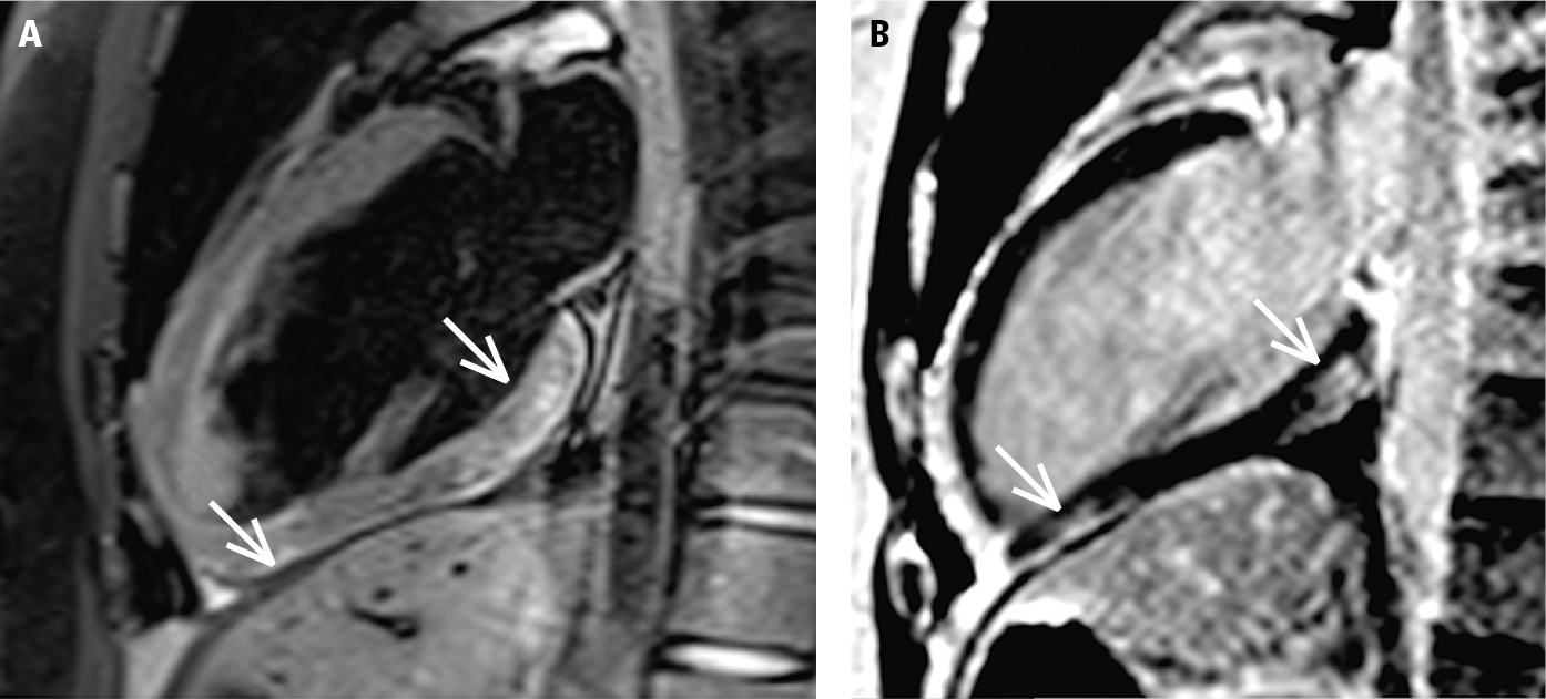
Cardiac magnetic resonance imaging (MRI) of a patient with suspected acute myocarditis, 2-chamber view. Both MRI criteria for acute myocarditis are met. A, features of myocardial edema (high signal intensity of the basal and apical inferior myocardial segments; arrows), the T2-dependent criterion. B, presence of the typical subepicardial late gadolinium enhancement in the area corresponding to the area of the edema (arrows), the T1-dependent criterion. Figure courtesy of Dr Łukasz Małek.