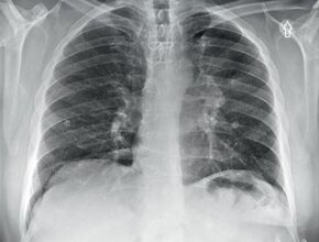
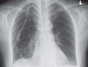
A 21-year-old male patient with a fever of unknown origin (FUO) and hemoptysis. You order a chest x‑ray and PA and lateral views are obtained (Fig. 1).
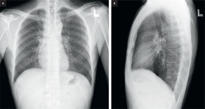
Figure 1. a, PA chest x-ray, b, lateral chest x-ray.
Question
What is the abnormality on the PA view and on the lateral view?
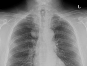
If we compare this patient’s PA chest x-ray (Fig. 1a) to a normal x-ray (Fig. 2) we can identify several abnormalities.
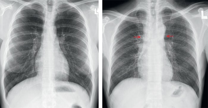
Figure 2. Normal PA chest x-ray for comparison.
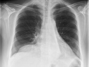
Figure 3. Chest X-ray PA. The upper mediastinum looks widened (indicated by the arrows)
and is whiter than normal.
First, the upper mediastinum looks widened (Fig. 3) and is whiter than normal. The right edge of the mediastinal border should create a straight border. In this case the right border is bulging outward (Fig. 4).
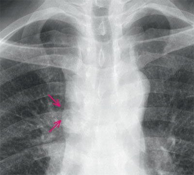
Figure 4. Chest x-ray, PA view (magnification). The right border of the mediastinum (indicated by the arrows) should be vertical and straight. In this case the border is bulging outward.
If you identified this abnormality then well done. To complete the interpretation of the chest x‑ray we need to go over all of the other structures to make sure we did not miss anything.
Are the trachea and the airways normal? YES
Are the lung fields normal? NO
Are the hila normal? YES
Is the heart normal? YES
Is the chest wall normal? YES
Is the pleura normal? YES
If you compare the appearance of the lungs with our normal x‑ray (Fig. 2) we can identify that the lungs are also abnormal. Within the lungs there are numerous, small, less than 3 mm nodules. These are non-calcified and appear to be distributed evenly throughout the lungs. The nodules are visible both on the PA film (Fig. 1a and 5) as well as on the lateral x‑ray (Fig. 1b and 6).
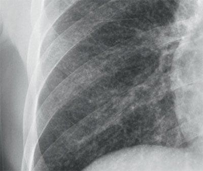
Figure 5. Chest x-ray, PA view (magnification). Numerous, small less than 3 mm nodules can be seen throughout both lungs.
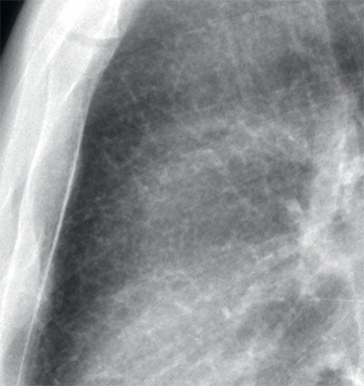
Ryc. 6. Chest x-ray, lateral view (magnification). Numerous, small less than 3 mm nodules can also be seen especially in the retrosternal air space.
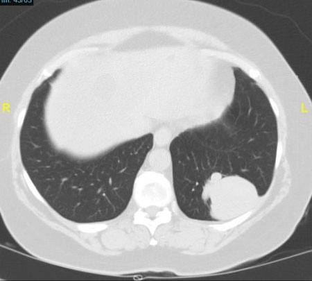
A subsequent computed tomography examination confirmed the presence of the nodules and the distribution (Fig. 7) as well as mediastinal adenopathy causing the widened mediastinum.
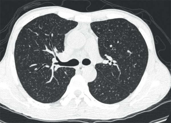
Ryc. 7. CT scan, axial image. Numerous, small, non-calcified, less than 3 mm nodules are seen throughout both lungs.
Summary
The chest x-ray shows numerous, small, non-calcified, less than 3 mm nodules throughout both lungs. In addition, there is widening of the mediastinum. Small lung nodules can be caused by infectious disease processes, such as tuberculosis, or by non-infectious processes, such as metastases or silicosis. For this reason, it is appropriate to have a differential diagnosis based on whether the patient is febrile or afebrile. If febrile, the differential diagnosis would include tuberculosis and histoplasmosis.
If afebrile, the differential diagnosis would also start with infectious causes, such as tuberculosis and histoplasmosis. However, you would then also include the following:
- Neoplastic causes – such as metastases from the thyroid gland.
- Occupational exposure – such as silicosis.
- Unknown etiology – such as sarcoidosis.
Diagnosis
Miliary Tuberculosis
This patient was a recent immigrant from Asia. The combination of the military pattern and adenopathy, strongly points to tuberculosis infection.
You can find more interesting cases in the app "Discover Radiology: Chest x-ray interpretation".
Price – only $6.99
References
1. Divinagracia R., Harris H.W.: Miliary tuberculosis. W: Schlossberg D., red.: Tuberculosis and nontuberculous mycobacterial Infections. 4th ed. Philadelphia, WB Saunders Company, 1999: 271–2842. Eisenhuber E., Mostbeck G., Bankier A. i wsp.: Radiologische Diagnostik der Lungentu berkulose. Radiologe, 2007; 47: 393–400