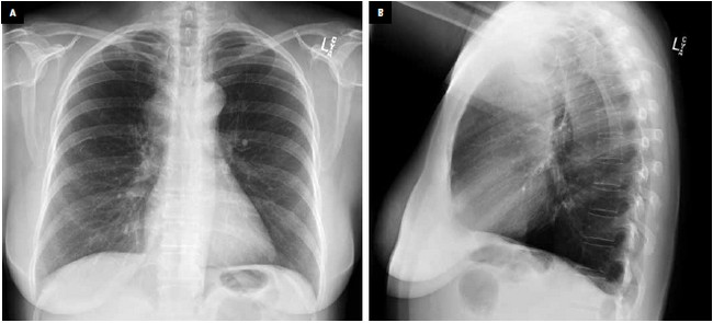
Figure 1. a, PA chest x-ray, b, lateral chest x-ray.
Can you identify an abnormality on the PA and lateral view?
If you identified the abnormality on the PA or lateral or both, you still need to complete your interpretation. You review all the other anatomical structures to make sure nothing else is wrong.
You should check
If you did not identify the abnormality on the PA or lateral, you will need to do a systematic review of the chest x-ray. My approach is to systematically evaluate the chest x-ray in the following 7 steps:1) Trachea and bronchi
2) Heart
3) Mediastinum
4) Hila
5) Lungs
6) Pleura and
7) Chest wall.
While doing a systematic evaluation of Fig. 1a and 1b you may be curious if something is wrong adjacent to the right side of the trachea. You would be correct.
What should a normal right paratracheal region look like on the x-ray (Fig. 2 and Fig. 3)?
Anatomy and radiological anatomy
The right paratracheal stripe represents the soft tissues adjacent to the right lateral border of the trachea and should measure less than 5 mm in thickness (Fig. 2 and Fig. 3).
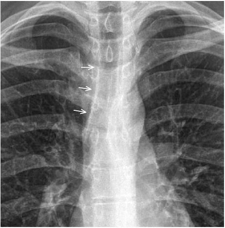
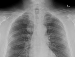
Figure 2. PA chest x-ray with white arrows pointing to the normal right paratracheal stripe.
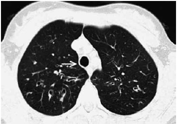
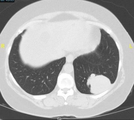
Figure 3. Axial CT scan. Normal right paratracheal stripe highlighted with white arrow (this patient has bronchiectasis).
Summary
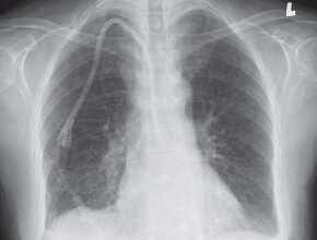
Now, if we look at this case again and compare it with a normal example, we can confidently see that the right paratracheal region is enlarged (Fig. 4). There is an opacity that bulges beyond the expected margin of the mediastinum (white arrows). This area is too wide and too white. This abnormality is not clearly identified on the lateral x-ray.
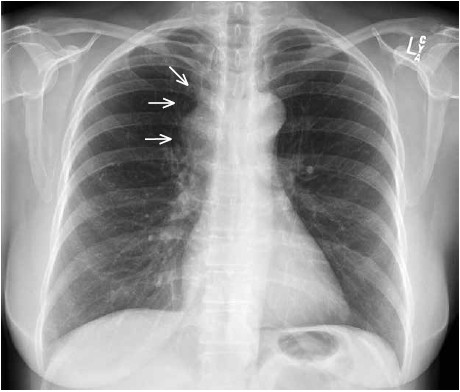
Figure 4. PA chest x-ray (the opacity of the lungs indicated by arrows).
Diagnosis
Enlarged right paratracheal stripe caused by sarcoid adenopathy
You can find more interesting cases in the app "Discover Radiology: Chest x-ray interpretation".
Price – only $6.99