 English
 Español
 українська

FiguresTop
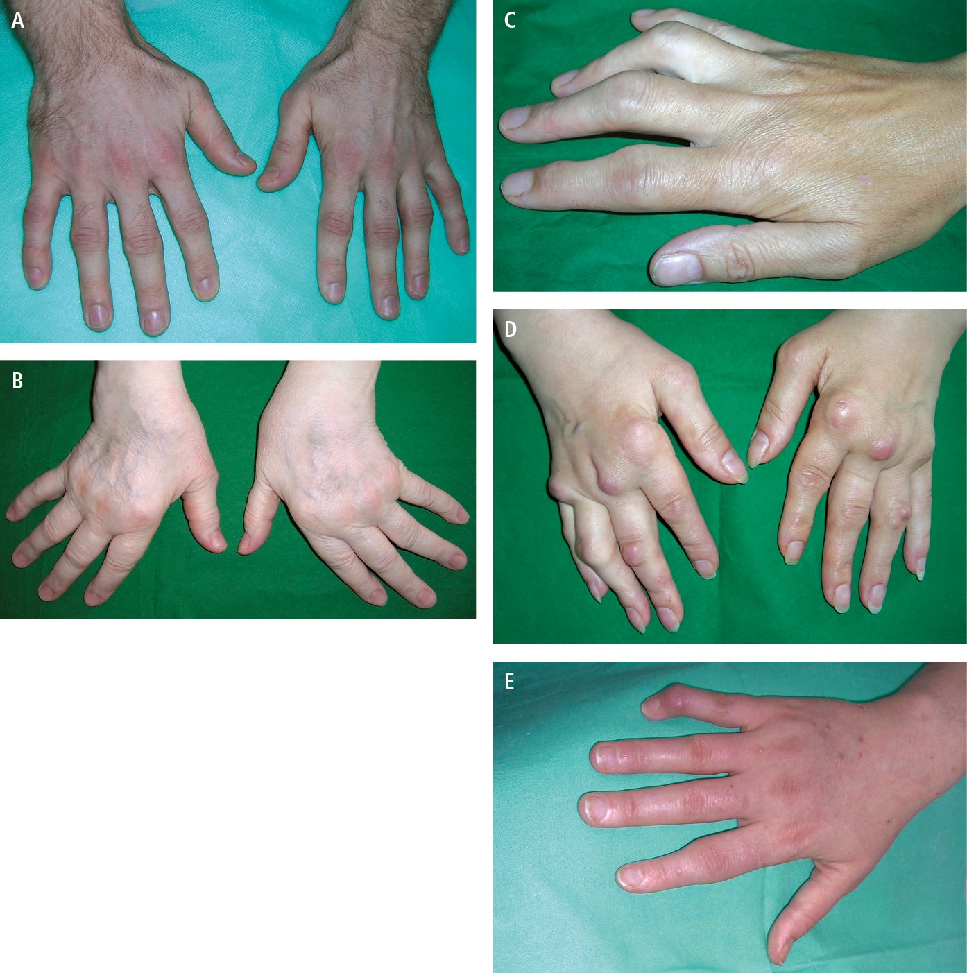
Figure 1.13-1. Rheumatoid arthritis. A, early changes: symmetric swelling of the metacarpophalangeal and proximal interphalangeal joints. B, ulnar deviation of the fingers and subluxation of the metacarpophalangeal joints. C, boutonnière deformity (fingers III and IV). D, swelling of the metacarpophalangeal and proximal interphalangeal joints, subluxation of the metacarpophalangeal joints, numerous rheumatoid nodules around the joints. E, swan-neck deformity of the little finger.
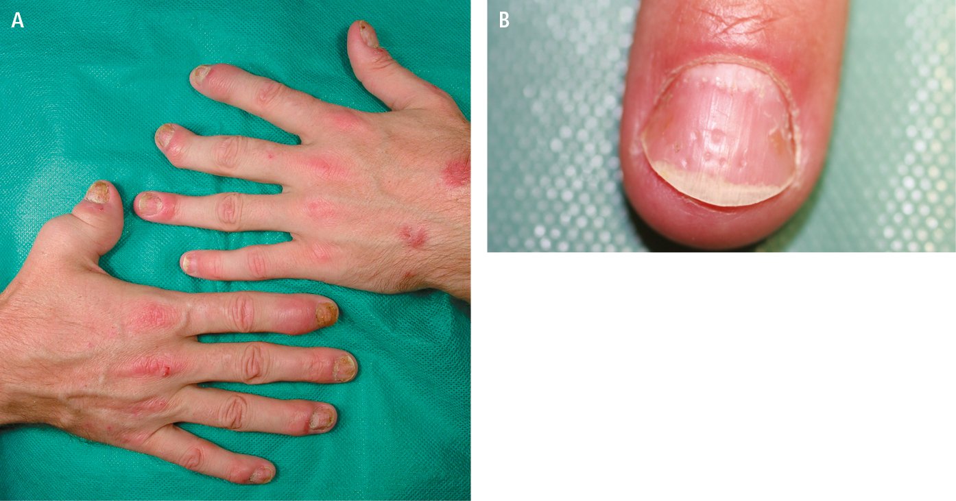
Figure 1.13-2. Psoriatic arthritis. A, psoriatic lesions on the skin of the outer aspect of both hands, telescopic finger I of the right hand, characteristic involvement of the distal interphalangeal joints, subluxation of the distal phalanx of finger III of the left hand, sausage-shaped finger II of the right hand. B, nail pitting.
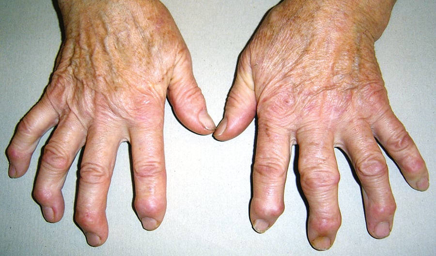
Figure 1.13-3. Osteoarthritis of the hand: Heberden nodes (over the distal interphalangeal joints) involving most fingers of both hands, a Bouchard node (over the proximal interphalangeal joint) on finger III of the left hand.
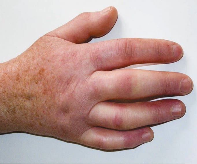
Figure 1.13-4. Systemic sclerosis. The skin on the digits is shiny, hardened, and limits full extension and full flexion of the fingers.
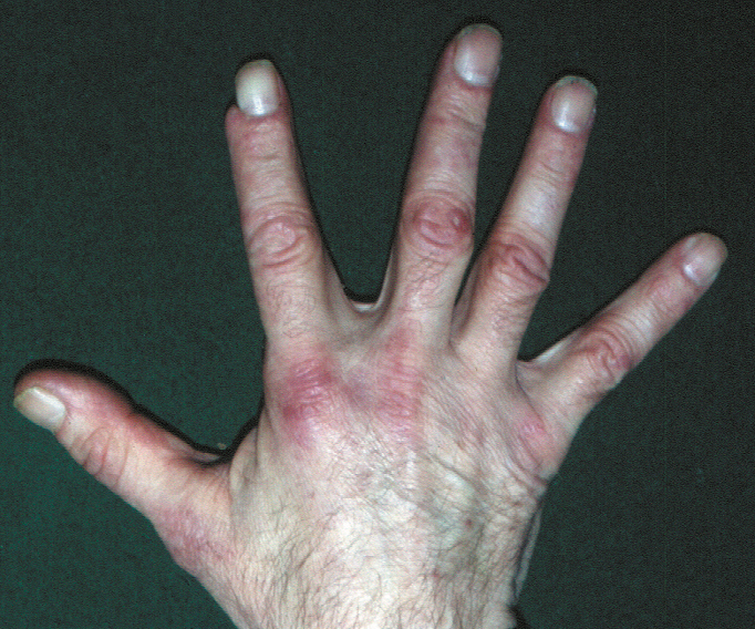
Figure 1.13-5. Polymyositis and dermatomyositis. Bluish papules over the interphalangeal and metacarpophalangeal joints (Gottron papules).
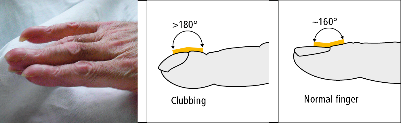
Figure 1.13-6. Clubbing.