 English
 Español
 українська

Bernard P, Antonicelli F. Bullous Pemphigoid: A Review of its Diagnosis, Associations and Treatment. Am J Clin Dermatol. 2017;18(4):513-528. doi:10.1007/s40257-017-0264-2. PMID: 28247089.
Venning VA, Taghipour K, Mohd Mustapa MF, Highet AS, Kirtschig G. British Association of Dermatologists’ guidelines for the management of bullous pemphigoid 2012. Br J Dermatol. 2012;167(6):1200-1214. doi:10.1111/bjd.12072. Epub 2012 Nov 2. PMID: 23121204.
Definition, Etiology, PathogenesisTop
Bullous pemphigoid (BP) is a chronic autoimmune skin disease characterized by generalized, pruritic, tense bullae. It is the most common autoimmune bullous disease and mainly affects elderly individuals, particularly patients aged >70 years. The hallmark of BP is the presence of circulating and tissue-bound autoantibodies against hemidesmosomal proteins BP180 (type XVII collagen or BPAG2) or less commonly against BP230 (BPAG1), which are structural components of the basement membrane zone at the dermo-epidermal junction. Approximately 85% to 90% of the antibodies target the juxtamembranous extracellular noncollagenous 16th A (NC16A) domain of BP180.
BP may be induced by drugs. Several agents have been implicated to cause drug-induced BP, including diuretics (furosemide, spironolactone) and angiotensin-converting enzyme inhibitors (captopril, enalapril, lisinopril). Newer classes of drugs have also been associated with the induction of BP, such as dipeptidyl peptidase–4 inhibitors (eg, linagliptin, sitagliptin) and programmed death–1 (PD-1) and programmed death ligand–1 (PD-L1) checkpoint inhibitors (eg, nivolumab).
Clinical Features and Natural HistoryTop
BP is characterized by widespread, tense vesicles and bullae on the background of normal-appearing or erythematous skin (Figure 4.1-1). The bullae usually contain clear, serous fluid, but may be hemorrhagic or blood tinged. Mucous membranes, particularly the oral mucosa, are involved in 10% to 30% of patients. In ~30% of patients the development of blisters can be preceded by a prodromal nonbullous stage. This stage could be either urticarial or manifested by eczematous lesions that persist for several weeks or months.
Several variants of BP have been reported, including localized, vulvar, acral, dyshidrotic, prurigo nodularis–like, vegetating, and nonbullous.
BP is associated with a higher prevalence and increased risk of neuropsychiatric disorders, including dementia, Parkinson disease, and stroke. Patients with active BP are also at increased risk of venous thromboembolism (deep vein thrombosis, pulmonary embolism) and strokes. The relation between BP and malignancy remains controversial. Some reports described an association with increased malignancy; however, there is insufficient evidence to justify extensive screening.
DiagnosisTop
The diagnosis of BP is based on the combination of clinical presentation, histologic findings, direct immunofluorescence findings, and detection of circulating autoantibodies.
1. Blood and immunologic tests: Circulating autoantibodies can be detected by indirect immunofluorescence (anti-skin antibodies in pemphigoid) in ~70% to 80% of patients with BP using the salt-split skin method. New techniques such as enzyme-linked immunosorbent assay (ELISA) are more specific and can be useful in monitoring disease activity.
It is also recommended to order a complete blood count, liver and kidney function tests, and serologic tests for hepatitis B and C in advance of potential initiation of systemic and immunosuppressive therapy. Eosinophilia is present in 30% to 50% of patients.
2. Skin biopsies: Two skin biopsies should be performed when BP is suspected. One lesional biopsy specimen from the edge of a newly formed, intact blister should be fixed in formaldehyde for standard histologic examination, and one perilesional biopsy specimen should be placed in Michel transport medium for direct immunofluorescence. Histopathology of lesional skin demonstrates a subepidermal blister with an eosinophil-rich infiltrate in the papillary dermis. Direct immunofluorescence of the perilesional skin is the gold standard to detect tissue-bound autoantibodies. It typically shows a linear deposition of IgG, C3, or both, at the basement membrane.
TreatmentTop
Treatment of BP depends on many factors such as age of the patient, severity and extent of the disease, and medical comorbidities. Prompt discontinuation of medication is recommended if drug-induced BP is suspected. Treatment options for BP: Table 4.1-1. Treatment with potent topical glucocorticoids can be initiated by a primary care physician, but systemic agents should be used under the supervision of a medical dermatologist.
Prognosis Top
Although some patients have self-limited disease over a few years, the majority have a chronic course with exacerbations and remissions.
Tables and FiguresTop
|
Drug |
Dosage |
Adverse effects |
Special considerations |
|
Superpotent topical glucocorticoids (0.05% clobetasol propionate cream or ointment) |
Topically on affected areas once daily or bid |
– Skin atrophy – Acne – Purpura – Hypertrichosis – Hypopigmentation |
Should be avoided if there is extensive BSA involvement (>10%) due to risk of systemic absorption. Should not be used continuously for >3 weeks |
|
Systemic glucocorticoids (prednisone) |
Prednisone 0.5-1 mg/kg/d PO until full resolution of itching and blisters, then tapered down slowly over a few months |
– Hyperglycemia – Hypertension – Peripheral edema – Osteoporosis – Myopathy – Cataracts – Glaucoma – Peptic ulcer disease – Leukocytosis – Neutrophilia – Lymphopenia – Insomnia – Irritability – Mood changes |
|
|
Tetracycline antibiotics (tetracycline, minocycline, doxycycline) |
Tetracycline 500 mg PO qid or minocycline 100 mg PO bid, or doxycycline 100 mg PO bid |
– GI upset – Photosensitivity – Drug-induced lupus with minocycline |
Can be given with or without nicotinamide (vitamin B3). Nicotinamide is an over-the-counter product and has been used at doses of 500-2000 mg/d due to a possible anti-inflammatory effect |
|
Azathioprine |
TPMT level must be checked prior to therapy initiation. The maximum doses are 2 mg/kg/d (TPMT >19 U), 1-1.5 mg/kg/d (TPMT, 13.7-19 U), and 0.5 mg/kg (TPMT, 5-13.7 U) |
– GI upset – Myelosuppression – Immunosuppression – Increased risk of infection – Increased risk of cancers including nonmelanoma skin cancers – Pancreatitis |
Requires follow-up monitoring of CBC, liver and kidney function tests |
|
MMP or MPA |
1-3 g/d (MMP) or 360-1080 mg bid (MPA) |
– GI upset – Myelosuppression – Immunosuppression – Increased risk of infection – Increased risk of cancers – Sterile pyuria – Dizziness |
Requires follow-up monitoring of CBC, liver and kidney function tests |
|
Methotrexate |
7.5-20 mg/wk PO or SC |
– Nausea, vomiting, and GI upset – Fatigue – Leukopenia – Alopecia – Mucositis – Elevated liver enzymes – Pneumonitis |
Requires follow-up monitoring of CBC, liver and kidney function tests |
|
Dapsone |
G6PD levels should be checked prior to initiating dapsone. The dose ranges from 25 to 100 mg/d PO |
– Hemolytic anemia – Methemoglobinemia – Agranulocytosis – Hypersensitivity reaction – Peripheral neuropathy |
|
|
IVIG |
2-3 g/kg every 4 weeks |
– Transfusion reactions – Transfusion-associated circulatory overload – Anaphylaxis in IgA-deficient patients |
– Very expensive – Premedication with acetaminophen (INN paracetamol) and antihistamines may prevent transfusion reaction or reduce its severity |
|
Rituximab |
375 mg/m2/wk IV × 4 doses or 1 g IV on days 0 and 15 |
– Infusion reactions (fever, headache, nausea, and pruritus) within the initial 0.5-2 h of the first infusion |
Premedication with acetaminophen (INN paracetamol) and antihistamines, usually with glucocorticoids PO, is commonly used to prevent infusion reactions |
|
BSA, body surface area; CBC, complete blood count; GI, gastrointestinal; G6PD, glucose-6-phosphate dehydrogenase; INN, international nonproprietary name; IVIG, intravenous immunoglobulin; MMP, mycophenolate mofetil; MPA, mycophenolic acid; PO, oral; SC, subcutaneous; TPMT, thiopurine methyltransferase. | |||
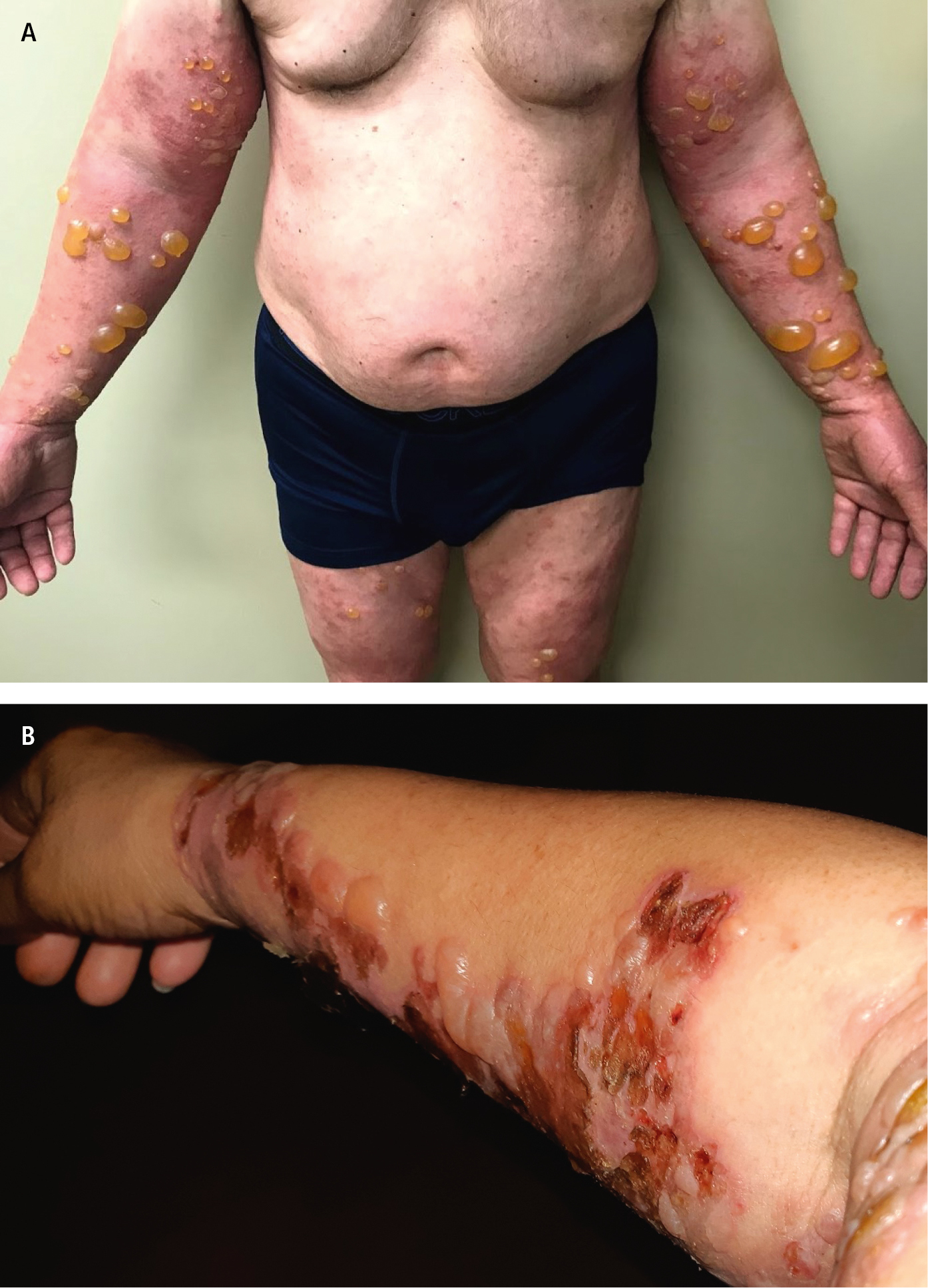
Figure 4.1-1. Tense bullae in bullous pemphigoid. A, on upper and lower extremities. B, on the right arm. Photograph courtesy of Dr Mohannad Abu-Hilal.