 English
 Español
 українська

Murrell DF, Peña S, Joly P, et al. Diagnosis and management of pemphigus: Recommendations of an international panel of experts. J Am Acad Dermatol. 2020;82(3):575-585.e1. doi:10.1016/j.jaad.2018.02.021. Epub 2018 Feb 10. PMID: 29438767; PMCID: PMC7313440.
Porro AM, Hans Filho G, Santi CG. Consensus on the treatment of autoimmune bullous dermatoses: pemphigus vulgaris and pemphigus foliaceus - Brazilian Society of Dermatology. An Bras Dermatol. 2019;94(2 Suppl 1):20-32. doi:10.1590/abd1806-4841.2019940206. Epub 2019 Jun 3. PMID: 31166407; PMCID: PMC6544031.
Harman KE, Brown D, Exton LS, et al. British Association of Dermatologists' guidelines for the management of pemphigus vulgaris 2017. Br J Dermatol. 2017;177(5):1170-1201. doi:10.1111/bjd.15930. PMID: 29192996.
Definition, Etiology, PathogenesisTop
Pemphigus refers to a group of rare autoimmune blistering diseases characterized by acantholysis (loss of cell-cell adhesion) and formation of intraepidermal blisters in the mucous membranes, skin, or both. Pemphigus vulgaris (PV) is the most common type, occurring in ~1 per 1,000,000 people per year. The hallmark of PV is the presence of circulating and tissue-bound autoantibodies against desmosomal proteins, desmoglein 1 (Dsg1), and desmoglein 3 (Dsg3), resulting in the loss of keratinocyte cell-cell adhesion within the epidermis.
Pemphigus can also be induced by drugs. Three groups of drugs have been implicated to cause the disease: thiol-containing drugs (eg, captopril and penicillamine), phenol-containing drugs (eg, rifampin, aspirin, and levodopa), and other drugs that do not contain thiols or phenols (eg, naproxen, diclofenac, verapamil, amlodipine, and lisinopril).
Clinical Features and Natural HistoryTop
There are 2 patterns of involvement in PV: mucosal and mucocutaneous. In the mucosal type, almost all patients develop painful mucosal erosions and ulcerations. The most commonly affected and often the first area to be involved is the oral cavity. Any area of the mouth can be affected, including the buccal mucosa, tongue, soft and hard palate, gingiva, and lips. Less commonly other mucosal surfaces may be involved, including the pharynx, larynx, nasal mucosa, esophagus, genital areas, vulva, and vagina. In the mucocutaneous type patients develop skin lesions in addition to mucosal erosions. Skin lesions appear as multiple flaccid blisters and erosions. Any area can be affected, but areas of predilection are the chest, back, face, and scalp (Figure 4.4-1). Unlike in bullous pemphigoid, lesions in PV are usually not itchy. Other variants of pemphigus: Table 4.4-1.
DiagnosisTop
The diagnosis of PV is based on the combination of clinical presentation, histologic findings, direct immunofluorescence findings, and detection of circulating autoantibodies.
1. Blood and immunologic tests: Circulating autoantibodies can be detected by indirect immunofluorescence (antiskin antibodies in pemphigus) in ~80% to 90% of patients with PV. There is a new technique, enzyme-linked immunosorbent assay (ELISA) for antibodies against Dsg1 and Dsg3, which is more specific and more sensitive and can be useful in monitoring the disease activity. It is also recommended to order complete blood count, liver and kidney function tests, and serologic tests for hepatitis B and C for possible initiation of systemic and immunosuppressive therapies.
2. Skin biopsies: Two skin biopsies should be performed when PV is suspected. One lesional biopsy specimen from the edge of a newly formed lesion should be fixed in formaldehyde for standard histologic examination, and 1 perilesional biopsy specimen should be placed in Michel transport medium for direct immunofluorescence.
Histopathology of the affected skin demonstrates suprabasal epidermal acantholysis and intraepidermal clefting with superficial mixed dermal inflammatory infiltrates including eosinophils. The base of the blister may have intact keratinocytes separated from one another, giving the “tombstone” appearance. Direct immunofluorescence of the perilesional skin is the gold standard to detect tissue-bound autoantibodies. It typically shows intercellular deposition of IgG, C3, or both, within the epidermis, giving the “chicken wire” appearance.
TreatmentTop
If not treated promptly, PV is associated with high mortality and morbidity, and therefore it should be managed by a medical dermatologist or a physician experienced in the treatment of autoimmune bullous diseases.
Treatment of PV depends on many factors such as age of the patient, severity and extent of the disease, and medical comorbidities. Medication should be promptly discontinued if drug-induced PV is suspected. Systemic glucocorticoids remain the gold standard treatment for PV. Available treatment options: Table 4.4-2.
Follow-upTop
Follow-up of a patient with PV involves monitoring the disease activity, adjusting therapy, and screening for adverse effects of immunosuppressive therapy. Once the disease activity is under control and healing is established, the dose of systemic glucocorticoids should be tapered over the following months. To avoid relapse, the tapering process should be paused if ongoing, active disease of the skin or mucosal surfaces is detected at any visit. If disease control is not achieved within several weeks of treatment with oral glucocorticoids or if relapse occurs, dosage of systemic glucocorticoids and use of adjuvant therapies should be re-evaluated. Serum titers for anti-Dsg1 and anti-Dsg3 antibodies can be used to monitor the disease activity.
PrognosisTop
Before the introduction of systemic glucocorticoids and adjuvant therapies, the mortality rate associated with PV ranged from 60% to 90%. With the currently available therapy, the mortality rate is ~10%, but it remains ~2-fold to 3-fold higher than in the general population. Complications of PV include sepsis and fluid/electrolyte imbalance, among others. Much of the morbidity and mortality associated with PV is related to the consequences of chronic systemic steroid use, such as increased risk of infection, hypertension, type 2 diabetes, and osteoporosis.
The disease course varies greatly among individuals. Remission can take months to years in both patients on therapy and those off therapy. Approximately 50% of patients with PV achieve complete remission off therapy after a mean treatment duration of 3 years. Adjuvant therapy, particularly the use of rituximab, can help induce remission and reduce the risk of relapse. Factors contributing to a poorer prognosis include advanced age, extensive disease, and greater initial severity.
Tables and FiguresTop
|
Pemphigus foliaceus (Figure 2) |
– No mucosal involvement – Usually milder skin lesions on the chest, back, face, and scalp |
|
Pemphigus herpetiformis |
– Characterized by grouped, pruritic, vesiculobullous lesions in herpetiform and annular arrangement – Mucosal involvement is uncommon |
|
Pemphigus vegetans (Figure 3) |
– Characterized by hyperkeratotic, verrucous, vegetative plaques, mostly affecting intertriginous areas – Involvement of oral mucosa is common |
|
IgA pemphigus (subcorneal pustular dermatosis and intraepidermal neutrophilic IgA dermatosis) |
Groups of vesicles and pustules on the erythematous background, mostly on the trunk, proximal extremities, and in intertriginous areas |
|
Paraneoplastic pemphigus |
– Severe, painful, hemorrhagic oral erosions and ulcerations extending to lips – Polymorphic skin lesions (eg, flaccid pemphigus-like blisters; tense pemphigoid-like bullae; erythema multiforme–like, graft-versus-host disease–like, and purple, lichen planus–like lesions) – Severe bronchiolitis (bronchiolitis obliterans) can also develop |
|
Drug |
Dosage |
Adverse effects |
Special considerations |
|
Superpotent topical glucocorticoids (0.05% clobetasol propionate cream or ointment) |
Topically on affected areas once daily or bid |
– Skin atrophy – Acne – Purpura – Hypertrichosis – Hypopigmentation |
|
|
Systemic glucocorticoids |
Prednisone 0.5-1 mg/kg/d PO until full resolution of itching and blisters, then tapered down slowly over several months |
– Hyperglycemia – Hypertension – Peripheral edema – Osteoporosis – Myopathy – Cataracts – Glaucoma – Peptic ulcer disease – Leukocytosis – Neutrophilia – Lymphopenia – Insomnia – Irritability – Mood changes |
|
|
Azathioprine |
TPMT level must be checked prior to therapy initiation. The maximum doses are 2 mg/kg/d (TPMT >19 U), 1-1.5 mg/kg/d (TPMT, 13.7-19 U), and 0.5 mg/kg/d (TPMT, 5-13.7 U) |
– GI upset – Myelosuppression – Immunosuppression – Increased risk of infection – Increased risk of cancers including nonmelanoma skin cancers – Pancreatitis |
Requires follow-up monitoring of CBC and liver and kidney function tests |
|
MMP or MPA |
1-3 g/d (MMP) or 360-1080 mg bid (MPA) |
– GI upset – Myelosuppression – Immunosuppression – Increased risk of infection – Increased risk of cancers – Sterile pyuria – Dizziness |
Requires follow-up monitoring of CBC and liver and kidney function tests |
|
Cyclophosphamide |
1-3 mg/kg/d PO |
– GI upset – Alopecia – Myelosuppression – Immunosuppression – Increased risk of infection – Infertility in both men (irreversible azoospermia) and women (irreversible amenorrhea) – Increased risk of malignancies – Hemorrhagic cystitis |
Used only in refractory cases |
|
IVIG |
2-3 g/kg every 4 weeks |
– Transfusion reactions – Transfusion-associated circulatory overload – Anaphylaxis in IgA-deficient patients |
– Very expensive – Premedication with acetaminophen (INN paracetamol) and antihistamines may prevent or reduce severity of transfusion reaction |
|
Rituximab |
375 mg/m2/wk IV for 4 doses or 1 g followed by another dose of 1 g after 2 weeks |
Infusion reactions (fever, headache, nausea, and pruritus) within the initial 0.5-2 h of first infusion |
Premedication with acetaminophen (INN paracetamol) and antihistamines, usually with glucocorticoids PO, is commonly prescribed to prevent infusion reactions |
|
CBC, complete blood count; GI, gastrointestinal; INN, international nonproprietary name; IVIG, intravenous immunoglobulin; MMP, mycophenolate mofetil; MPA, mycophenolic acid; PO, oral; TPMT, thiopurine methyltransferase. | |||
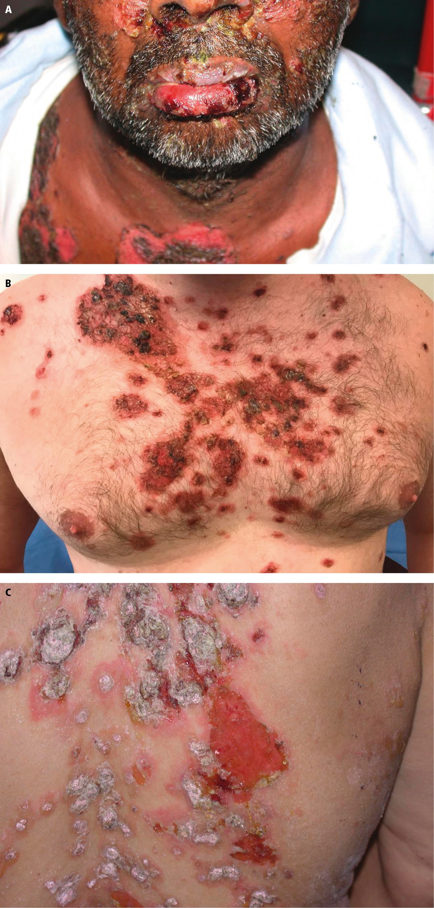
Figure 4.4-1. Pemphigus vulgaris. A, diffuse facial and lip erosions and crusted plaques. B, extensive flaccid blisters and erosions on the chest. C, diffuse erosions and plaques on the back. Photograph courtesy of Dr Scott Walsh.
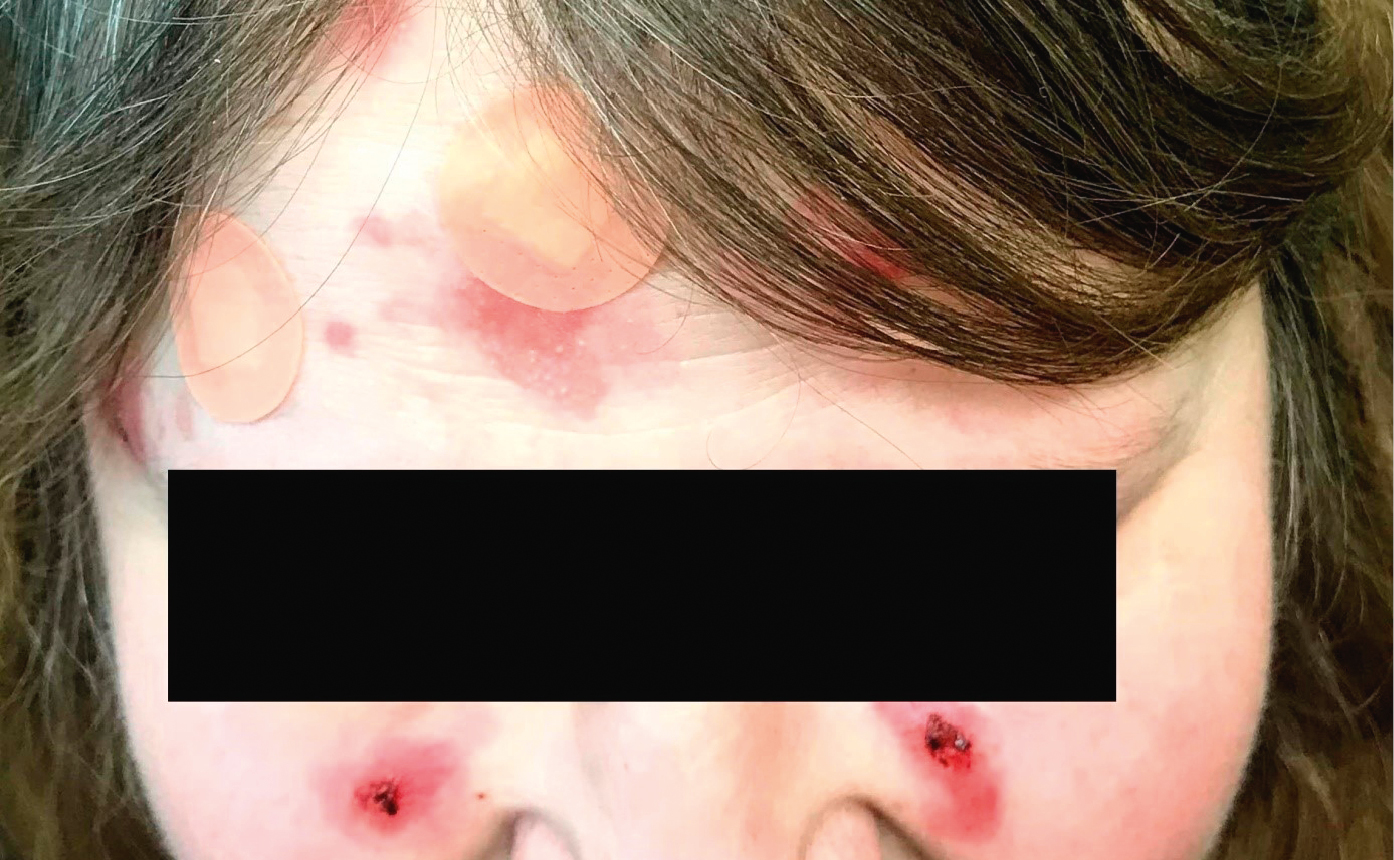
Figure 4.4-2. Pemphigus foliaceus. Superficial flaccid erosions and crusted plaques on the face without oral involvement. Photograph courtesy of Dr Mohannad Abu-Hilal.
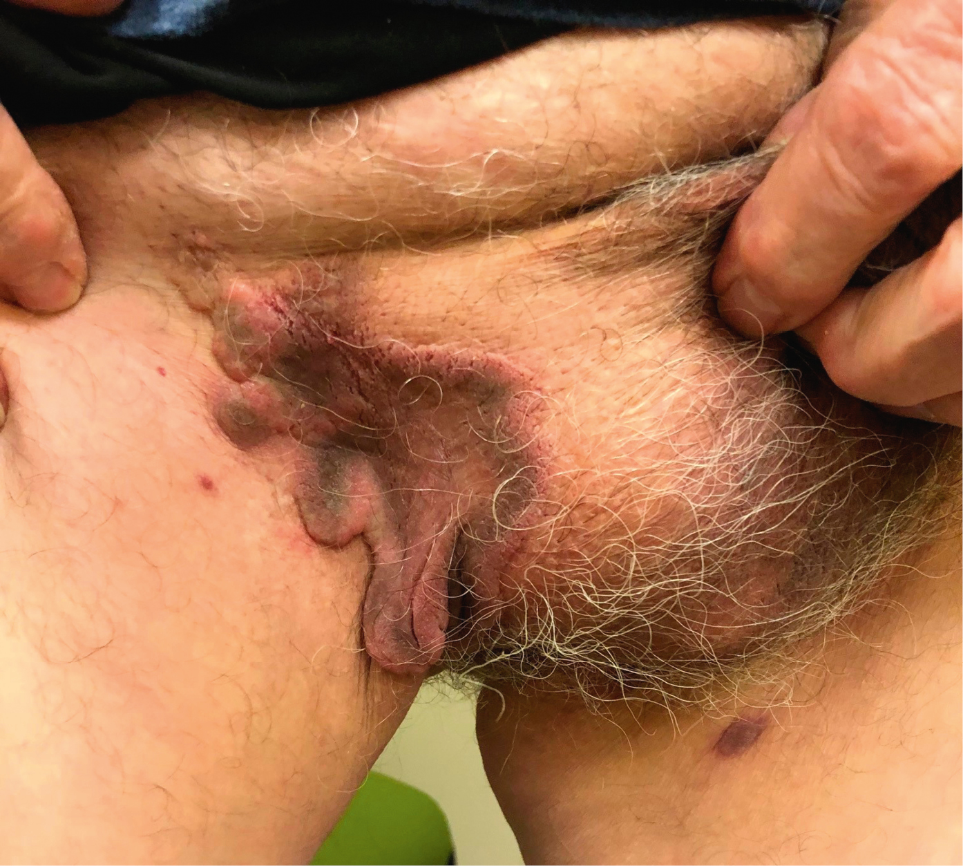
Figure 4.4-3. Pemphigus vegetans. Cauliflower-like vegetating plaques in the groin (flexural areas). Photograph courtesy of Dr Mohannad Abu-Hilal.