English
 Español
 українська

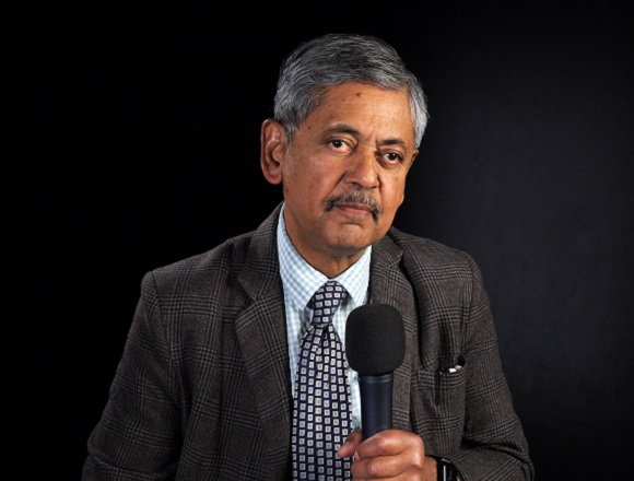
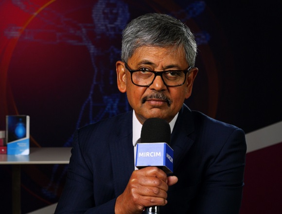
Bhaskar Dasgupta, MB, BS, MD, is a consultant rheumatologist and head of rheumatology at Southend University Hospital, UK. He developed guidelines on polymyalgia rheumatica, giant cell arteritis, and the concept of point-of-care rheumatology ultrasound.
Has temporal artery ultrasonography replaced biopsy in patients with suspected giant cell arteritis (GCA)?
Absolutely. We have the European Alliance of Associations for Rheumatology (EULAR) recommendations for imaging in large-vessel vasculitis and they clearly mention that if you have suggestive clinical symptomatology—and I was talking today about the Southend GCA Probability Score—so, the Southend GCA Probability Score will give you an idea whether GCA is likely, highly probable, unlikely, less probable, or intermediate.
In these patients, we do eight-vessel ultrasonography. We look at the temporal arteries, the frontal parietal branches, and the axillary arteries, and together with the pretest probability derived from the Southend GCA Probability Score and the temporal artery ultrasonography, we can come to a very nice posttest probability of GCA, where the GCA is likely, GCA is unlikely, or GCA is uncertain.
Only if we find after the ultrasonography that the GCA diagnosis still remains uncertain, that’s the time we do a temporal artery biopsy. So, overall, the number and the frequency of temporal artery biopsies have gone down dramatically. It’s ~10% to 15% in our fast-track GCA clinic.