 English
 Español
 українська

Stevens SM, Woller SC, Kreuziger LB, et al. Antithrombotic Therapy for VTE Disease: Second Update of the CHEST Guideline and Expert Panel Report. Chest. 2021 Dec;160(6):e545-e608. doi: 10.1016/j.chest.2021.07.055. Epub 2021 Aug 2. Erratum in: Chest. 2022 Jul;162(1):269. doi: 10.1016/j.chest.2022.05.028. PMID: 34352278.
Definition, Etiology, PathogenesisTop
Chronic venous insufficiency is venous stasis due to a retrograde flow of blood, stenosis, or occlusion of the veins. Chronic venous insufficiency may be caused by varicose veins (persistent dilation of a superficial vein ≥3 mm in diameter measured in a standing position), postthrombotic syndrome, primary venous valve incompetence, venous compression syndromes (eg, popliteal entrapment syndrome), and rarely by congenital conditions such as the Klippel-Trénaunay syndrome.
Venous insufficiency in the pelvic (ovarian, perineal, periuterine, gluteal) veins may contribute to a condition referred to as pelvic congestion syndrome (PCS), which is associated with chronic pelvic pain. The pathophysiology of PCS is not fully understood but may relate to pelvic vein reflux and varicosities. There are no established diagnostic criteria and there is uncertainty as to the effectiveness of various proposed treatment modalities, which typically involve vascular interventions, surgical interventions, or both.
Risk factors: Age >40 years, female sex, genetic factors causing weakness of the vein walls and valves (causing so-called primary varicose veins), pregnancy, working in sitting or standing positions, obesity. Regardless of the cause, the primary factor leading to the development of chronic venous insufficiency is venous hypertension, which may result from agenesis, hypoplasia, incompetence, or damage of the venous valves, as well as from venous occlusion or stenosis caused by thrombosis (and lack of or incomplete recanalization following a thrombotic event) or by compression of the veins.
In some cases, venous ulcers are accompanied by stasis dermatitis, which may be caused by trauma, microtrauma, bacterial infection, or contact allergy.
Clinical FeaturesTop
1. Symptoms: Patients with early venous insufficiency complain of a feeling of “heaviness” and “fullness” in the lower extremities, which usually worsens in the evening and improves after resting with elevated legs. Other symptoms include visible blue-colored, dilated superficial veins, painful muscle cramps in the calf (particularly at night), as well as restless leg syndrome. In the more advanced stages patients usually develop dull pain that worsens during the day; pain while walking (called venous claudication), which is rarely observed in patients with venous insufficiency, indicates occlusion of deep veins of the lower leg.
2. Signs:
1) Telangiectasias (Figure 3.19-1) (dilated intradermal venules <1 mm in diameter, small spider veins, reticular varicose veins) that with time progress to wide tortuous varicose veins of the long (great) saphenous vein (Figure 3.19-2) and short (small) saphenous vein.
2) Edema (in early disease it is pitting, reversible, and resolves after overnight rest; later it becomes hard, nonpitting, and irreversible).
3) Rusty-brown discoloration of the skin of the lower leg (Figure 3.19-3).
4) Venous ulcers (Figure 3.19-4) (typically above the medial malleolus; advanced ulcers involve the entire circumference of the lower leg).
5) Dry or oozing eczema of varying severity and persistent cellulitis (frequent in patients with advanced chronic venous insufficiency).
6) Lipodermatosclerosis.
7) Secondary lymphedema.
8) Atrophie blanche (white atrophy): A pattern of healing in the area of poor blood supply (Figure 3.19-5).
3. Signs and symptoms of stasis dermatitis include severe erythema and inflammatory lesions on one or both lower extremities, in some cases with a generalized hematogenous reaction (associated with erythematous or fine papular rash that frequently includes the skin of the head, trunk, and upper extremities) and severe pruritus. Bacterial superinfections of cutaneous lesions are frequent.
DiagnosisTop
Diagnosis is based primarily on the clinical manifestations and, secondarily, on the results of color Doppler ultrasonography of the veins in the lower extremity.
Bilateral or unilateral edema of the lower extremities (see Edema).
TreatmentTop
1. General recommendations: Avoidance of exposure to high temperatures and intense sunlight, avoidance of prolonged standing or prolonged sitting with the knee and hip joints flexed at right angles; maintaining an ergonomic workplace, including a chair with reclining backrest and a footstool; several minutes of walking or limb exercises after longer periods of sitting; regular recreational exercise (walking, jogging, cycling, swimming); frequent rests with the lower extremities raised above the heart level, supported along the entire length of the lower leg (not by a few isolated support points).
2. Compression therapy is the only method that can slow down the development of chronic venous insufficiency. It may also be used prophylactically in patients with early manifestations of venous insufficiency. Therapy may involve the use of compression bandages (in patients with venous ulcerations), compression stockings (selected on an individual basis to fit the limb after edema has improved or resolved: Table 3.19-8), and intermittent pneumatic compression (see Deep Vein Thrombosis).
Contraindications: Acute cellulitis, exudative skin diseases, Fontaine class III/IV arterial ischemia (ankle-brachial index [ABI] <0.6; before applying any type of compression, always examine the pulse in the lower extremities and measure ABI), limb deformities or severe arthritis that make it impossible to precisely apply appropriate pressure.
3. Pharmacologic treatment is an addition to (but never a replacement of) compression therapy. Natural or synthetic flavonoids (rutoside and its derivatives, hesperidin, diosmin), saponins (aescin), calcium dobesilate, and grape seed or citrus extracts may improve quality of life and alleviate symptoms in some patients, but they do not prevent the progression of the disease. The best evidence of effectiveness exists for saponins.Evidence 1Low Quality of Evidence (low confidence that we know true effects of intervention). Quality of Evidence lowered due to methodological limitations, imprecision, and indirectness (short duration of treatment of a life-long disease). Pittler MH, Ernst E. Horse chestnut seed extract for chronic venous insufficiency. Cochrane Database Syst Rev. 2012 Nov 14;11:CD003230. doi: 10.1002/14651858.CD003230.pub4. Review. PMID: 23152216.
1. Elevation of the limbs while sitting or lying down.
2. Compression therapy: Multilayer compression using special bandages or commercial multilayer compression systems for treatment of ulcers (recommended pressures are 40 mm Hg at ankle level and 17-20 mm Hg at popliteal level; maximum pressures in patients with mixed arteriovenous ulcers and patients with ABI 0.6-0.9 are 17-25 mm Hg).
3. Removal of necrotic tissues, wound debridement, transplant of cutaneous and/or musculocutaneous flaps.
4. Treatment of infection: Topical disinfectants containing octenidine, dressings with 7% to 10% iodinated povidone or with ethacridine solution, and systemic (not topical!) antibiotics.
5. Treatment of pain is particularly important during debridement and dressing changes.
6. Treatment of stasis dermatitis: Oral antihistamines, topical glucocorticoids, and compresses of 1% tannin and 0.1% silver nitrate.
7. Correction of possible protein malnutrition impairing the healing process (assessment of the patient’s nutritional status is necessary before starting treatment of venous ulcers).
8. Patients with ulcers that have not healed for >3 months despite appropriate treatment should be referred for a specialist consultation. Malignant lesions within the ulcer must be excluded.
1. Indications: Severe symptoms of chronic venous insufficiency, complications of varicose veins (inflammation, rupture, bleeding, trophic skin lesions, venous ulcers), cosmetic reasons. Do not refer patients with occlusion of the deep veins for surgical treatment.
2. Methods: Stripping of varicose veins, open surgical treatment of incompetent perforators (Linton method), minimally invasive surgery (microphlebectomy, cryosurgery, laser surgery), sclerotherapy (obliteration of the veins by injecting sclerotic agents). Recurrences of varicose veins after surgery are frequent (affecting up to 50% of patients). Good long-term effects of surgery largely depend on the continued use of compression treatment.
Tables and FiguresTop
|
Class |
Pressurea |
Indications |
|
I |
20-30 |
Prevention of venous thrombosis, prophylaxis of thrombosis and varicose veins in pregnant patients, small varicose veins during pregnancy, complaints of heavy and tired legs, small varicose veins with no visible edema, postsurgical treatment of varicose veins |
|
II |
30-40 |
Large varicose veins during pregnancy, varicose veins with mild edema, history of superficial vein phlebitis, prior sclerotherapy of varicose veins, healed small ulcers |
|
III |
40-50 |
Very large varicose veins with significant edema, successfully healed large ulcers, posttraumatic edema, reversible lymphedema |
|
IV |
50-60 |
Severe postthrombotic syndrome, irreversible lymphedema |
|
a Pressure at ankle level (mm Hg). | ||
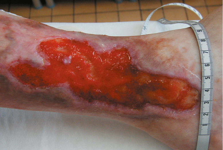
Figure 3.19-4. Venous ulcerations. Figure courtesy of Dr Leszek Masłowski.