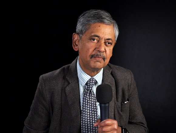
Bhaskar Dasgupta, MD, is a professor of rheumatology at Southend University Hospital, UK, and honorary professor of Essex University, UK.
How to differentiate giant cell arteritis (GCA) from Takayasu arteritis?
This is very important, particularly in countries where you get a high incidence of Takayasu disease. In many ways they share a common pathogenesis. They are both large-vessel vasculitides, but, of course, Takayasu occurs in countries where you don’t see the cranial type of GCA. For example, you see Takayasu in Japan, you see Takayasu in India, in many other parts of the world where you do not see the typical cranial GCA.
The way to differentiate the two: one is age. Takayasu is usually a condition that will occur in the younger age group. We have a definition: generally it occurs below the age of 40 or at least below the age of 50. Takayasu patients have a more chronic onset of disease, whereas GCA has a more acute onset of disease. Takayasu usually does not cause cranial manifestations, so you do not usually get the headache or jaw pain. The visual symptoms that you get in Takayasu are due to ischemia of the carotids or the vertebrals because of involvement by Takayasu.
On the other hand, Takayasu causes some specific changes, which GCA does not cause. For example, it causes renal artery stenosis. You get abdominal involvement and you get secondary hypertension due to renal artery stenosis. You get hypertension, you can also get pulmonary artery involvement in Takayasu disease.
There are many ways they are similar, but there are ways they are dissimilar. One particular dissimilarity is the involvement of the subclavians. Takayasu commonly involves the subclavian [artery], whereas GCA does not usually involve the subclavian [artery].
The bottom line is we should be imaging all patients, whether it be GCA or Takayasu, to make sure that the vascular territories are uninvolved and we should pick them up at the first sign of any involvement.
 English
 Español
 українська

